


 返回

Original article link: HeYuan Bio – Chemical Genetics Technology
Chemical genetics (also known as pharmacogenetics) is an important emerging technology that has emerged alongside optogenetics in recent years. By engineering certain biomolecules to enable them to interact with small molecules they previously could not recognize, this approach allows for the controlled and reversible regulation of biomolecular activity—compounds can be added or removed at will to initiate or terminate specific biochemical reactions. This technique has already found broad applications in areas such as signal transduction, drug development, and functional genomics.
They are widely used to noninvasively enhance or suppress neuronal activity with cell specificity. Although DREADDs lack the precise temporal control afforded by optogenetics, they are well suited for applications that require long‑term modulation of neural circuits—precisely the kind of manipulation often needed in therapeutic settings. Moreover, many FDA‑approved drugs target GPCRs, and since DREADDs are engineered GPCRs, they hold considerable promise for drug discovery and development.
Currently, the biomolecules that have been engineered include nucleic acid hybrids, protein kinases, various metabolic enzymes, and G protein–coupled receptors (GPCRs). Numerous chemogenetic platforms based on GPCRs have now been developed, such as allele-specific activation of genetically encoded receptors, first established in 1991; receptors activated solely by synthetic ligands (RASSLs), introduced in 1998; engineered receptors; and designer receptors exclusively activated by designer drugs (DREADDs), which emerged in 2007. Among these, DREADDs has become the most widely used chemogenetic technique, and this review will focus primarily on DREADDs.
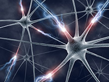
Figure 1. Chemical genetics techniques
1) DREADDs technology
DREADD technology was developed by Bryan L. Roth and colleagues, who engineered the structure of a G-protein–coupled receptor—the acetylcholine receptor—so that it can be selectively activated or inhibited only by a specific compound, clozapine-N-oxide (CNO). These modified receptors differentially modulate distinct GPCR signaling cascades, including Gq, Gi, Gs, Golf, and β‑arrestin pathways; among them, Gq‑DREADDs and Gi‑DREADDs are the most widely used. When these receptors are expressed in cells, their downstream effects vary depending on the presence of CNO.
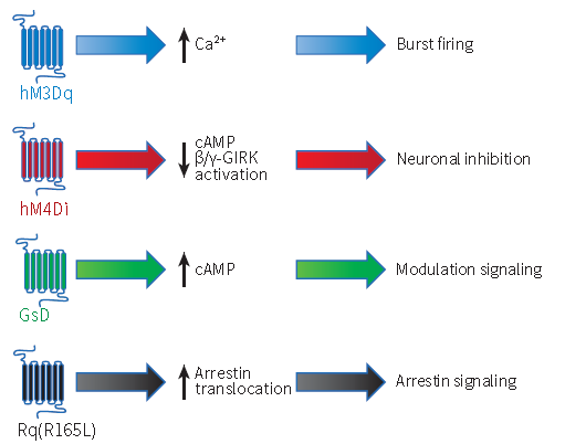
Figure 2. Mechanisms of action of several commonly used DREADD receptors (Scott M. Sternson & Bryan L. Roth, Annu. Rev. Neurosci., 2014)
2) Common DREADD receptors
1. Gq-DREADDs and hM3Dq:
The original Gq‑DREADD, hM3Dq, was engineered by modifying the human muscarinic acetylcholine receptor subtype M3 (also known as hM3). Under normal physiological conditions, hM3 binds acetylcholine and then couples with Gq‑type G proteins, activating the phospholipase C–inositol trisphosphate–intracellular calcium signaling pathway (Figure 3).
Surprisingly, mutating just two residues—Y3.33C and A5.46—renders the hM3 receptor incapable of coupling to acetylcholine while allowing it to bind CNO at nanomolar concentrations. This engineered hM3 receptor is designated hM3Dq (human M3 muscarinic DREADD receptor coupled to Gq). Because Y3.33 and A5.46 are conserved across different human muscarinic acetylcholine receptor subtypes, M1 and M5 have also been successfully converted into Gq‑DREADDs (hM1Dq and hM5Dq). Nevertheless, to date, hM3Dq remains the most widely used Gq‑DREADD.
In different cell types, CNO‑induced activation of hM3Dq yields distinct outcomes. For example: 1) In mature neurons, CNO‑induced hM3Dq leads to neuronal depolarization and enhanced excitability—this is the most commonly exploited function of hM3Dq, namely, promoting neuronal firing; 2) In astrocytes, some studies have reported that CNO‑induced hM3Dq increases intracellular Ca²⁺ release, thereby modulating autonomic nervous system–related physiological processes; 3) Beyond the nervous system, other investigations have demonstrated that, for instance, in pancreatic β cells, acute CNO treatment enhances insulin secretion, whereas chronic CNO exposure results in an increase in β‑cell numbers; in hepatocytes, hM3Dq activation elevates blood glucose levels, possibly due to increased glycogenolysis and gluconeogenesis.
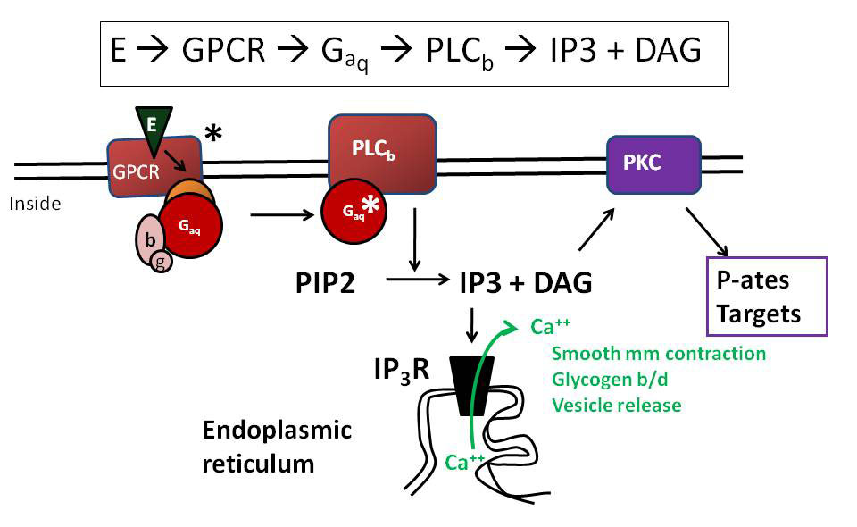
Figure 3. hM3 binds acetylcholine and, as a signaling molecule, activates Gq‑coupled GPCR signaling pathways.
2. Gi-DREADDs and hM4Di:
Y3.33 and A5.46 are conserved across different muscarinic acetylcholine receptor subtypes; accordingly, researchers can also mutate the Y3.33 and A5.46 residues in M2 and M4 mAChRs. Since M2 and M4 activate Gi‑coupled channels downstream, this yields Gi‑DREADDs, designated hM2Di and hM4Di, which can engage Gi‑mediated signaling pathways (Figure 4).
Gi‑coupled GPCRs can activate G‑protein‑gated inwardly rectifying potassium channels (GIRKs). Upon CNO administration, activation of hM2Di and hM4Di receptors suppresses neuronal firing; among these, hM4Di is the most widely used Gi‑DREADD. Additionally, studies have shown that hM4Di can inhibit neurotransmitter release, thereby dampening neuronal activity.
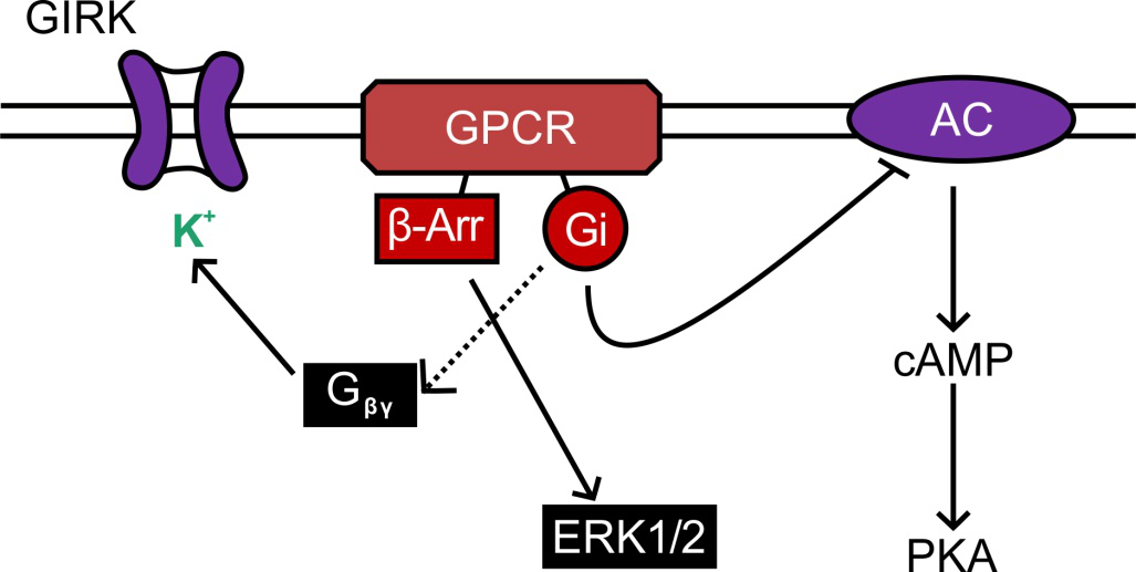
Figure 4: hM4 binds to acetylcholine and, as a signaling molecule, activates Gi‑coupled GPCR signaling pathways.
For more chemogenetic tools and viruses, please search...
3) Application Strategies of DREADD Technology
The application of DREADD technology using viral vectors typically involves the following key steps (Figure 5):
1. Based on the experimental objective, select an appropriate DREADD receptor: generally, hM3Dq is used to activate neurons, while hM4Di is used to inhibit neurons.
2. Utilize viral vectors to express DREADD receptors in vivo.
3. Design an experimental protocol to administer the CNO drug to animals within an appropriate time window to activate the receptor; administration routes include intracerebral stereotactic injection, intraperitoneal injection, and oral gavage.
4. Phenotypic analysis, which assesses changes resulting from alterations in neuronal activity using behavioral or electrophysiological approaches.
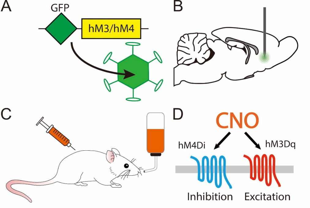
Figure 5. General strategy of DREADD technology
4) Technical Advantages of DREADDs
DREADD and optogenetic techniques are very similar in their applications, and they share the same underlying goals. So, how should we choose between them in our everyday research? The criterion for selection lies in an analysis of the respective advantages and disadvantages of each technique:
The main advantages of DREADD technology are:
1. The experimental requirements are minimal, and the procedure is straightforward: unlike optogenetic techniques, which necessitate fiber optics, laser controllers, and other specialized equipment, DREADD technology relies solely on standard pharmacological methods—simply injecting or administering CNO.
2. Non-invasive: Unlike optogenetic techniques, which require craniotomy and implantation of optical fibers, this approach does not impose additional weight that could alter mouse behavior, enabling the modulation of activity in specific brain regions and neuronal populations while the mouse remains fully free to move.
3. Achieving long-term activation or inhibition of neuronal activity: Due to their distinct mechanisms, optogenetic techniques rely on the opening of light‑sensitive ion channels, which requires ion flux across the cell membrane to generate membrane potential changes and modulate neuronal activity. In contrast, sustained ion transport against concentration gradients—such as that mediated by ion pumps—consumes substantial ATP, potentially leading to cellular damage and death; moreover, the thermal effects of light stimulation can also harm cells. By comparison, DREADDs express a receptor that can sustainably activate or inhibit neuronal activity for several hours without disrupting normal cellular physiology.
4. High safety: CNO is a metabolite of clozapine, an FDA‑approved drug, and its in vivo use is relatively safe. Moreover, many FDA‑approved drugs target GPCRs, whereas DREADDs are engineered GPCRs; thus, DREADDs may offer substantial potential in drug development.
Table 1: Comparison of the Advantages and Disadvantages of Optogenetics and Chemogenetics
Optogenetics technology DREADDs technology
In terms of temporal precision, the technique achieves millisecond‑level or even sub‑millisecond resolution, and can sustain neuronal activation or inhibition for several hours without disrupting normal cellular physiology.
Spatially, by employing brain‑targeted injections, cell‑type‑specific promoters, and subcellular organelle‑targeting peptides, light‑sensitive proteins can be precisely localized to specific cells or organelles, enabling manipulation at the single‑cell level. Similarly, DREADD receptors can be selectively expressed in a particular cell type through targeted injection and the use of cell‑type‑specific promoters.
Operational technique: Craniotomy-based implantation of optical fibers requires optical fibers, a laser controller, and other equipment, making the procedure relatively challenging. In contrast, the non-invasive approach involves only standard pharmacological techniques—simply injecting or administering CNO orally.
Case Studies of DREADD Technology Applications
1. hM4Di
① The client published an article: Science. (IF=41.058). Mu D, et al. (2017). A central neural circuit for itch sensation. [Adeno-associated virus, itch, optogenetics, chemogenetics]
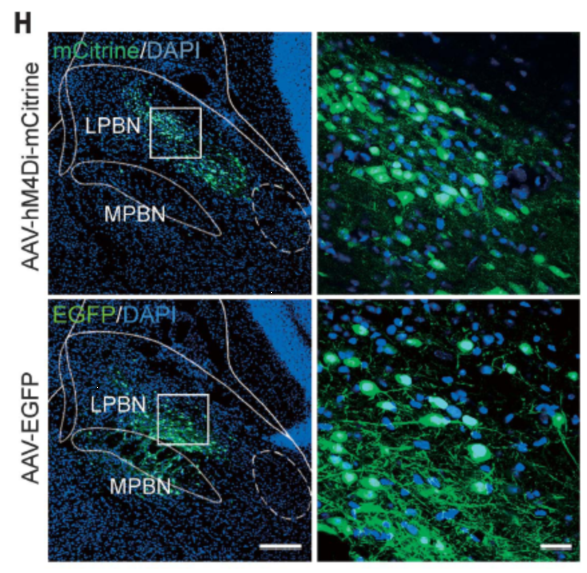
Injection site: mouse PBN
Vector: AAV-hSyn-HA-hM4Di-IRES-mCitrine
Serotype: AAV2/9
Viral titer: 1.0 × 10¹³ VG/mL
Injection volume: 150 nl
Observation period: 3 weeks
② Client-published article: Neuron. (IF=14.319). Xu HF, et al. (2019) A Disinhibitory Microcircuit Mediates Conditioned Social Fear in the Prefrontal Cortex. [Social anxiety disorder, AAV, chemogenetics]
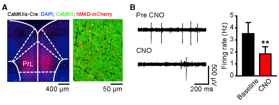
Injection site: mouse PrL
Vector: AAV2/9-hSyn-DIO-hM4D(Gi)-mCherry
Serotype: AAV2/9
Viral titer: 3.44 × 10¹³ VG/mL
Injection volume: 100–200 nl
Observation period: 4 weeks
 Telephone consultation


