


 返回

Original article link: HeYuan Bio – Exosomes
Exosomes are a type of extracellular vesicle (EV) secreted by cells, with sizes ranging from 30 to 150 nm. They possess a bilayer membrane structure and a cup‑shaped morphology, containing a rich array of cargo—including nucleic acids, proteins, and lipids—and mediating intercellular molecular communication. Exosomes are widely present in cell culture supernatants and various bodily fluids, such as blood, lymph, saliva, urine, semen, and milk, and they are also found in tissue samples, including brain, muscle, and adipose tissues.
Brief description of the brain tissue isolation procedure: The brain tissue is cut into thin slices, transferred to a centrifuge tube, and treated with a digestion solution. After incubation in a water bath and repeated gentle pipetting up and down, the sample is subjected to intermittent, slow aspiration and dispensing until digestion is complete. Next, culture medium is added to the digestion mixture, the suspension is thoroughly mixed, and the sample is kept on ice. A series of differential ultracentrifugation steps follows, including impurity removal, filtration through a membrane, and ultracentrifugation. Finally, the exosomes are resuspended in PBS, and the resulting suspension is used for subsequent characterization by transmission electron microscopy (TEM), nanoparticle tracking analysis (NTA), and marker‑based Western blotting.

Figure 1: Extracellular vesicle secretion
All cells are capable of secreting exosomes, yet the exosomes secreted by different cell types vary considerably in both quantity and cargo, which in turn determines the distinct functions they perform. Exosomes play a broad role in intercellular transport of biomolecules and signaling, thereby regulating cellular physiological processes. Moreover, they contribute to antigen presentation, immune evasion, induction of normal cell transformation, and promotion of tumorigenesis and metastasis. In addition, exosomes can serve as “natural nanoparticles” for drug delivery.
What are the databases related to exosomes?
The exoRBase database collects and characterizes all long RNAs present in human blood-derived exosomes, including circRNAs, lncRNAs, and mRNAs.
The EVpedia and Vesiclepedia databases compile information on proteins, mRNAs, miRNAs, lipids, and other molecules identified in various vesicle studies.
The ExoCarta database primarily catalogs 286 research studies across several species, including humans, rats, mice, and sheep, covering information on proteins, mRNAs, miRNAs, lipids, and more.
Sample Preprocessing, Exosome Isolation, and Storage:
Exosomes are present in various bodily fluids of humans and animals, allowing researchers to select different sample sources for exosome‑related studies. As exosomes originate from multivesicular bodies—membrane‑bound vesicles formed by invagination of the plasma membrane—and are distributed within the extracellular matrix, obtaining high‑purity exosomes requires the effective removal of all cellular debris and other unwanted impurities.
1) Cell culture supernatant 2) Plasma/Serum
3) Urine 4) Cerebrospinal fluid
5) Follicular fluid 6) Endometrial fluid
7) Bile 8) Amniotic fluid
9) Ascites 10) Pleural fluid
Different experimental sample types require specific precautions during collection; for example, cell culture supernatants must be harvested in exosome‑free serum, plasma samples should not be collected in heparin‑anticoagulated tubes, and urine collections necessitate the addition of a bacteriostatic agent.
Various methods for isolating exosomes from biological fluids have been developed, primarily leveraging their size, density, and immunological characteristics. Obtaining highly purified exosomes is a critical step for subsequent exosome research; to date, differential ultracentrifugation is widely recognized as the “gold standard” for exosome isolation and remains the method of choice in high‑impact publications.
Extracellular vesicles can be stored at 4°C for short-term use (within one week). For long-term storage, they should be kept at −20°C or −80°C. Alternatively, the vesicles can be aliquoted and stored separately at −20°C or −80°C.
Exosome detection methods:
Following isolation, exosomes must undergo a series of characterization steps to confirm their identity. These characterization methods span multiple dimensions, ranging from physical properties to surface molecular markers.
l Transmission electron microscopy (TEM): This method is suitable for examining the ultrastructure of the double-layered membrane of exosomes, which typically appears as a cup-shaped or hemispherical structure with a depression on one side.
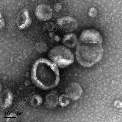
Figure 2 Transmission electron microscope
l Nanoparticle Tracking Analysis: abbreviated as NTA, this method preserves the native state of exosomes, offers rapid detection, and provides information on exosome particle size and concentration.
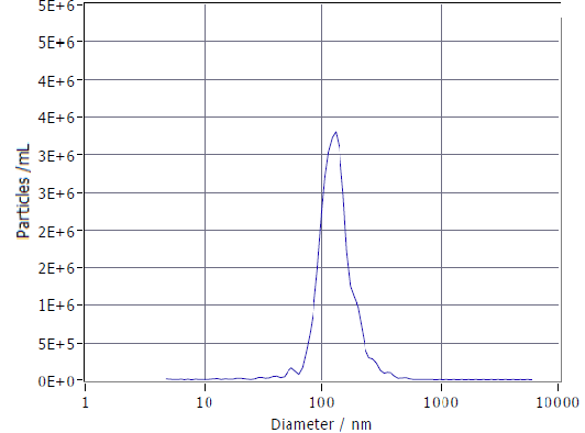
Figure 3 NTA
l Western blot analysis of molecular markers: Exosome marker proteins include tetraspanin family members such as CD9, CD63, and CD81; cytoplasmic proteins like actin and annexins; molecules involved in biological functions, such as ALIX (apoptosis‑related protein X), TSG101 (tumor susceptibility gene 101), heat shock proteins (HSP70, HSP90), as well as cell‑secreted specific proteins.
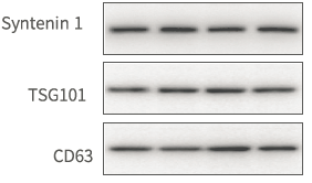
Figure 4: Western Blot (Tian Su et al., ACS Nano, 2019)
High-throughput exosome analysis
Exosomes contain proteins and nucleic acids that reflect their cellular origin, enabling the transport of proteins, mRNA, miRNA, lncRNA, circRNA, and other molecules into recipient cells, thereby participating in intercellular communication. The protein and RNA profiles of exosomes derived from different cell types vary, making them promising biomarkers for the early diagnosis of various diseases and potential carriers for targeted therapeutics.
1. High-throughput miRNA sequencing
2. mRNA high-throughput sequencing
3. lncRNA microarray (human, mouse)
4. ceRNA microarray (human, mouse)
5. Proteomic Analysis (iTRAQ, TMT, Label-Free)
Exosome labeling or tracing:
l Lipophilic dye–labeled exosomes: In currently published studies on exosomes, most researchers employ lipophilic dyes for labeling, with widespread applications both in vivo and in vitro. Lipophilic dyes are broadly categorized into two main types; the first includes PKH67 (green fluorescence) and PKH26 (red fluorescence). Because these dyes can stably associate with the lipid bilayer of exosomes, they provide robust staining and are widely used.
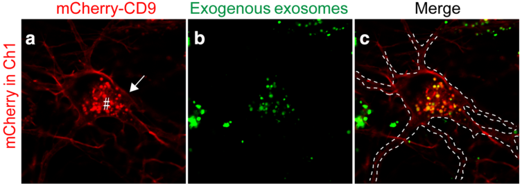
Figure 5: Interaction between PKH67-labeled exosomes and neurons (Juan Carlos Polanco et al., Acta Neuropathol Commun. 2018)
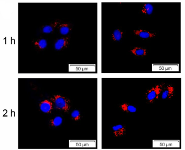
Figure 6. Co-culture of PKH26-labeled exosomes with MDA-MB-231 cells (Mengyu Yu et al., Cancer Sci. 2019)
The second category comprises lipophilic dyes of the Di series, including DiI (orange fluorescence), DiO (green fluorescence), DiD (red fluorescence), and DiR (deep red fluorescence). Notably, DiR’s near-infrared fluorescence can penetrate cells and tissues, making it suitable for in vivo imaging and tracking.
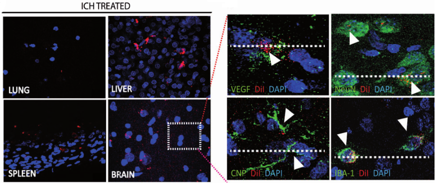
Figure 7. In vivo distribution of DiI-labeled exosomes following intravenous injection, as observed in various organs (Laura Otero-Ortega et al., J Cereb Blood Flow Metab. 2018).
l Lentivirus-mediated CD63–GFP expression: The expression cassettes for the exosomal protein CD63 and the green fluorescent protein GFP are cloned into a plasmid, which is then packaged into lentiviral particles. These lentiviruses are subsequently used to infect cells, resulting in the secretion of exosomes that carry green fluorescence.
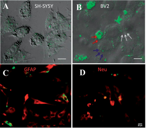
Figure 8. Exosomes labeled with GFP were co-cultured with SH-SY5Y, BV2, and DRG cells, respectively (Rui Ren et al., Artif Cells Nanomed Biotechnol. 2019).
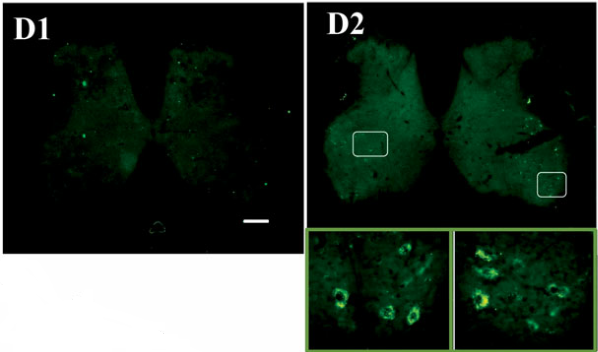
Figure 9. Fluorescence observed on day 1 (D1) and day 5 (D2) after injection of exosomes labeled with CD63‑GFP (Rui Ren et al., Artif Cells Nanomed Biotechnol. 2019).
Exosome Function Research:
Add the labeled exosomes to the recipient cells’ culture medium and co‑culture them, then assess changes in cellular functions such as proliferation, migration and invasion, and apoptosis. Alternatively, inject the exosomes into an animal model to observe phenotypic alterations and measure relevant physiological parameters.
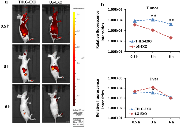
Figure 10: Intravenous injection of DiR-labeled exosomes into a mouse model of colorectal cancer (Gaofeng Liang et al., J Nanobiotechnology. 2020)
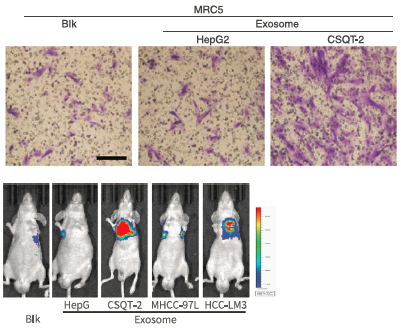
Figure 11: Functional studies of exosomes (Tian Fang et al., Nat Commun. 2018)
Our Advantages:
1) The experimental period is short;
2) Competitive pricing;
3) Comprehensive after-sales service;
4) Extensive project experience with an almost 100% success rate;
5) A complete suite of experimental services.
Our services:
1) Differential ultracentrifugation;
2) Transmission electron microscopy (TEM);
3) Nanoparticle Tracking Analysis (NTA);
4) Western blot analysis of protein markers;
5) Exosome high-throughput sequencing/chip;
6) Exosome labeling or tracing;
7) Exosome-mediated cellular function assays;
8) Exosome injection in animals;
9) Validation in animal models, among other approaches.
 Telephone consultation


