


 返回

Original article link: HeYuan Bio – Tumor Animal Models
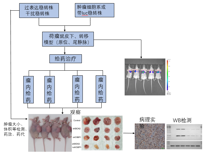
Tumor animal models play a crucial role in basic research on tumorigenesis, tumor progression, and metastasis, as well as in drug development and therapeutic strategies; therefore, the establishment of animal models is indispensable for cancer research. The commonly used tumor models in research include subcutaneous tumor formation, orthotopic tumor formation, and tail vein injection models.
Subcutaneous tumor model
A tumor animal model is established by directly implanting tumor cells (or tumor tissue) beneath the skin of mice. This model serves as a simple yet essential tool for preclinical evaluation of in vivo drug efficacy and also plays a critical role in studies of tumor pathogenesis and drug mechanisms of action.
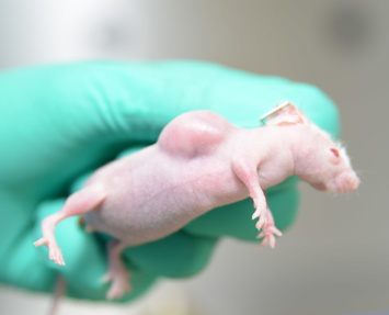
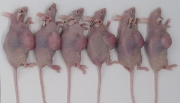
Subcutaneous tumorigenesis
This model is simple and convenient to operate, allowing for intuitive observation of tumor growth and facilitating the measurement of key parameters such as animal body weight, tumor growth curves, and tumor weights.
Orthotopic transplantation model
Orthotopic transplantation refers to the implantation of tumor cells or tumor tissue fragments directly into the tissues and organs of immunodeficient animals, thereby inducing tumor formation and the development of spontaneous metastatic foci.
The method involves directly implanting diluted tumor cells beneath the serosa of an organ. Alternatively, tumor cells may first be implanted subcutaneously until the tumor reaches a volume of approximately 500–800 mm³; the tumor tissue is then excised and cut into 1–3 mm³ fragments, which are subsequently implanted beneath the organ’s serosa, followed by closure of the serosal layer. This approach can also establish an orthotopic xenograft tumor.
Common sites for orthotopic inoculation include the brain, liver, muscle, and mammary fat pad.
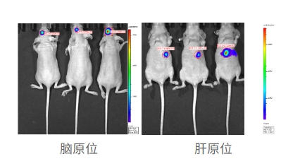
Orthotopic transplantation models more accurately mimic the in vivo microenvironment in which tumor cells grow, replicating both tumor growth and metastasis.
Tumor metastasis model via tail vein injection
Following intravenous injection via the tail vein, tumor cells first traverse the pulmonary capillary network and enter the arterial circulation, potentially giving rise to multiple metastatic foci throughout the body. However, due to their increased viscosity and propensity to aggregate, these cells are often trapped in the microvasculature of the mouse lung, predominantly resulting in pulmonary metastases; over time, they may also seed distant organs. This model is primarily used to establish tumor metastasis (hematogenous dissemination) models, hematologic malignancy models, or models of tumor lung metastasis.
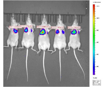
Intravenous injection of breast cancer cells into the tail vein
Tumor metastasis is the leading cause of cancer-related mortality in clinical practice; however, its mechanisms and underlying processes remain highly complex and incompletely understood. Establishing metastatic models enables more accurate simulation of tumor initiation and progression in humans, thereby enhancing the predictive value of drug screening and development.
The Shanghai Animal Experiment Platform of HeYuan offers a range of tumor‑bearing animal models, including subcutaneous xenografts, orthotopic xenografts, and tail‑vein injection–induced metastatic models, along with services such as histopathological sectioning, pharmacodynamic evaluation, and small‑animal imaging.
 Telephone consultation


