


 返回

Original article link: HeYuan Bio – Circuit Tracing
The circuitry connecting neurons is highly complex; fully and accurately mapping the structure and dynamics of these circuits is of paramount importance for elucidating the etiologies of numerous diseases and for developing novel therapeutic strategies.
Traditional neural tracing methods, such as electron microscopy, Golgi staining, dyes, and peptide-based markers, can reveal neuronal morphology and projections. A few tracers, including WGA and TTC, even enable transsynaptic labeling. However, these approaches suffer from drawbacks such as indirect signaling, lack of directional specificity, and substantial signal attenuation across synapses. Most critically, these dyes cannot deliver genetic material, limiting them to visualizing only protein–protein interactions. Since the 1980s, when Kristensson first employed the herpes simplex virus (HSV) for transsynaptic neuronal labeling, a growing array of viral vectors has increasingly gained prominence in circuit‑level tracing.
However, it should be noted that although these neurotropic viruses exhibit strong infectivity and specificity for labeling neural circuits, they are highly immunogenic; animals often die within a few days after injection, precluding behavioral assays or physiological studies. Consequently, they are currently best suited for identifying previously unknown neural circuits. Moreover, such viruses require biosafety containment at Level 2 or higher. As a result, an increasing number of studies now employ AAV for circuit‑tracing and labeling. Because AAV lacks the ability to replicate and elicits minimal immune responses, it does not cause significant toxicity nor disrupt normal animal physiology, making AAV the most widely used viral vector for circuit tracing.
Neurons are polarized cells: information is transmitted from the axon of an upstream neuron to the dendrites of a downstream neuron via synapses, followed by integration of incoming signals at the dendrites and soma, and then re‑emitted through the axon. Consequently, labeling of neural circuits involves two fundamental considerations: directionality (anterograde: labeling from soma to axon; retrograde: labeling from axon to soma) and whether the label crosses synapses (Figure 1).
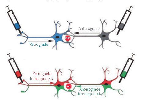
Figure 1. Objectives of neural circuit tracing techniques (adapted from Peter L. Strick et al., eLS, 2011)
Loop Tracing Application Case Studies
1. AAV-based anterograde non-cross-synaptic labeling
① The client published an article: Science. (IF=41.058). Mu D, et al. (2017). A central neural circuit for itch sensation. [Adeno-associated virus, itch, optogenetics, chemogenetics]
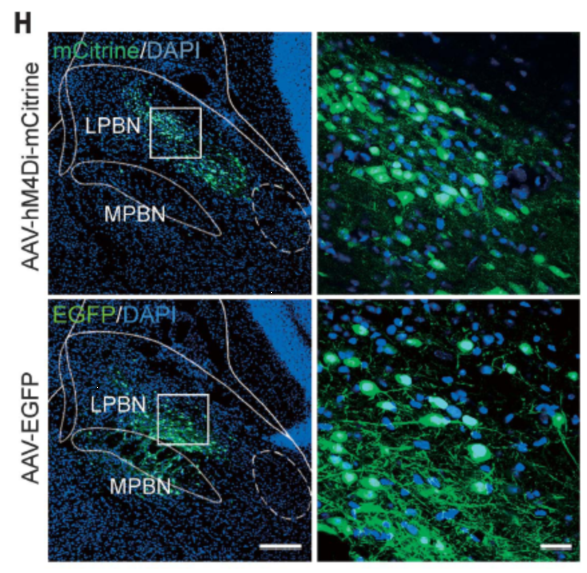
Injection site: mouse PBN
Vector: AAV-hSyn-HA-hM4Di-IRES-mCitrine
Serotype: AAV2/9
Viral titer: 1.0 × 10¹³ VG/mL
Injection volume: 150 nl
Observation period: 3 weeks
② Client-published article: Neuron. (IF=14.319). Tian-Le Xu, et al. (2019). Central Processing of Itch in the Midbrain Reward Center. [AAV, itch, optogenetics]
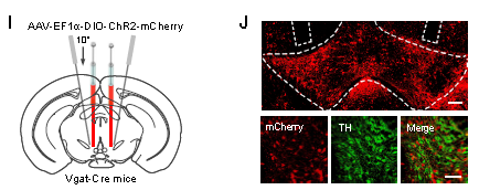
Injection site: VTA of Vgat-Cre mice
Vector: AAV-EF1a-DIO-ChR2(H134R)-mCherry
Serotype: AAV2/8
Viral titer: 7.39×10^12 VG/mL
Injection volume: 300–400 nl
Observation period: 2–3 weeks
2. AAV-based retrograde non-cross-synaptic labeling—rAAV2-retro
① Client-published article: Biological Psychiatry. (IF=11.984). Bing Xing Pan, et al. (2018). Chronic stress causes projection-specific adaptation of amygdala neurons via SK channel downregulation. [Adeno-associated virus, anxiety disorder, optogenetics]
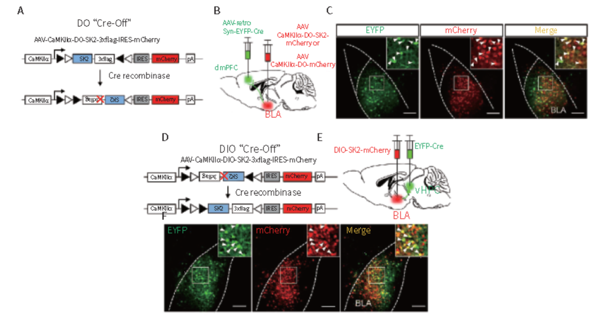
Injection site: mouse dmPFC, VHPC
Vectors: AAV2/1-retro-Syn-eYFP-Cre, AAV2/8-CaMKIIα-DO-SK2, and AAV2/8-CaMKIIα-DIO-SK2-mCherry
Injection volume: 300 nl
Observation period: 4 weeks
② Internal Test Results
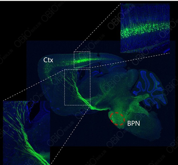
Injection site: Mouse BPN
Vector: rAAV2-retro-hSyn-EYFP
Observation period: 4 weeks
3. AAV-based transsynaptic labeling
3.1 AAV-Based Anterograde Transsynaptic Labeling—rAAV2/1
① The client published an article in Nature Neuroscience (IF = 19.912): Yao J, et al. (2018). A corticopontine circuit for initiation of urination. [Adeno-associated virus, neural circuit]
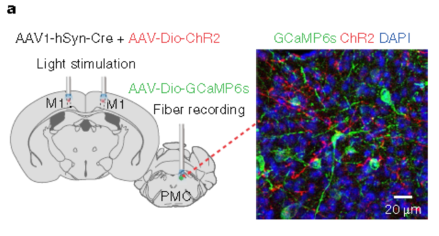
Injection site: mouse cortical M1 and PMC
Vectors: rAAV2/1-hSyn-Cre & rAAV2/9-DIO-hChR2(H134R)-mCherry, rAAV2/9-DIO-GCaMP6s
Viral titers: rAAV2/1: 5 × 10^12 VG/mL; rAAV2/9-DIO-hChR2: 1.2 × 10^13 VG/mL; rAAV2/9-DIO-GCaMP6s: 0.5 × 10^12 VG/mL
Injection volume: Multi-point injection, 30–40 nL per site.
Observation period: 4 weeks
② Client-published article: Science Advances. (IF=11.511). Ping Zheng, et al. (2019). Crucial role of feedback signals from prelimbic cortex to basolateral amygdala in the retrieval of morphine withdrawal memory. [Adeno-associated virus, addiction, AAV1, AAV-WGA-Cre]
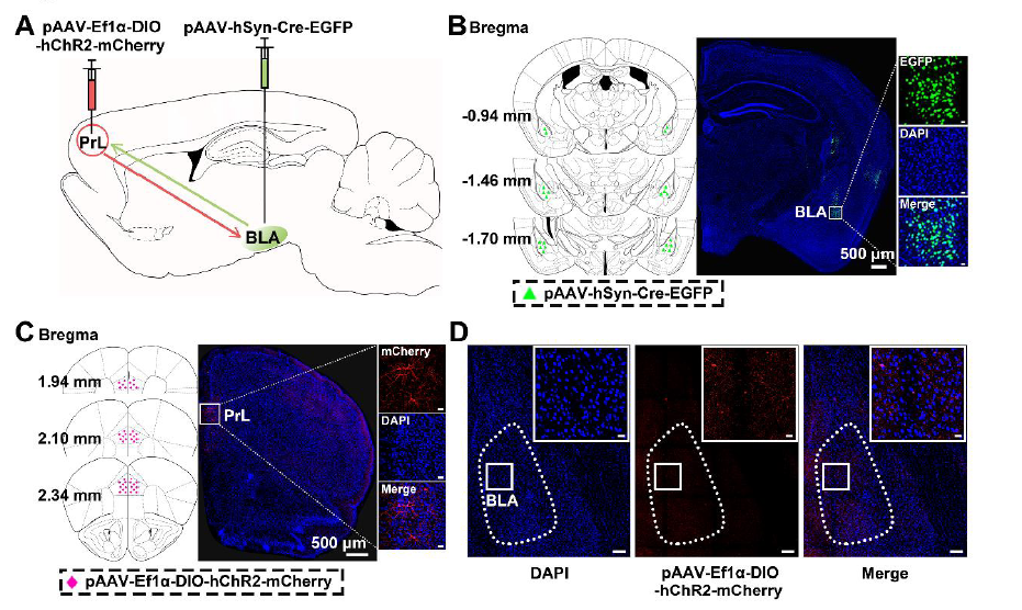
Injection site: mouse cortical BLA
Vector: rAAV2/1-hSyn-Cre-EGFP
Viral titer: 1.13 × 10^13 VG/mL
Injection volume: 60 nl
Observation period: 5 weeks
3.2 Cross-Synaptic Labeling Using WGA—AAV-WGA-Cre
Customer-published article: Science Advances. (IF=11.511). Ping Zheng, et al. (2019). Crucial role of feedback signals from the prelimbic cortex to the basolateral amygdala in the retrieval of morphine withdrawal memory. [Adeno-associated virus, addiction, AAV1, AAV-WGA-Cre]
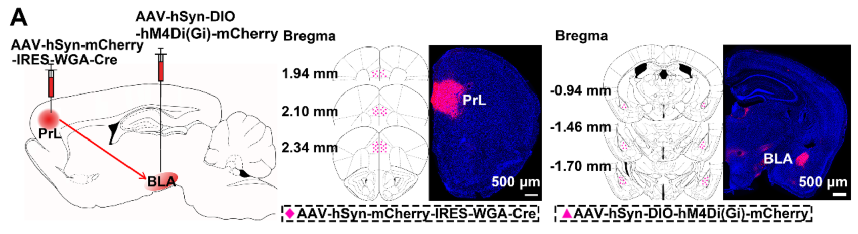
Injection site: mouse PrL
Vector: AAV-hSyn-mCherry-IRES-WGA-Cre
Viral titer: 4.28 × 10^12 VG/mL
Injection volume: 300 nl
Observation period: 4 weeks
 Telephone consultation


