


 返回

Original article link: HeYuan Bio – Adenovirus ADV
Adenovirus (ADV) is a non-enveloped, linear double-stranded DNA virus with broad tropism for various cell types and tissues. It induces a robust infection and is well suited for experiments requiring high‑level gene expression within a short time frame. Compared with other viral vectors, adenoviruses offer the advantage of accommodating longer insert fragments and exhibiting strong transcriptional activity.
In vitro, adenoviral vectors exhibit high transgene‑delivery efficiency—approaching 100% transduction—and can transduce a wide variety of cell types, making them an excellent tool for gene delivery to many cell types that are difficult to transfect. However, in vivo, due to their strong immunogenicity and robust infectivity, adenoviruses often elicit local tissue inflammation and systemic immune responses, which can compromise animal health parameters and the objectivity of experimental outcomes.
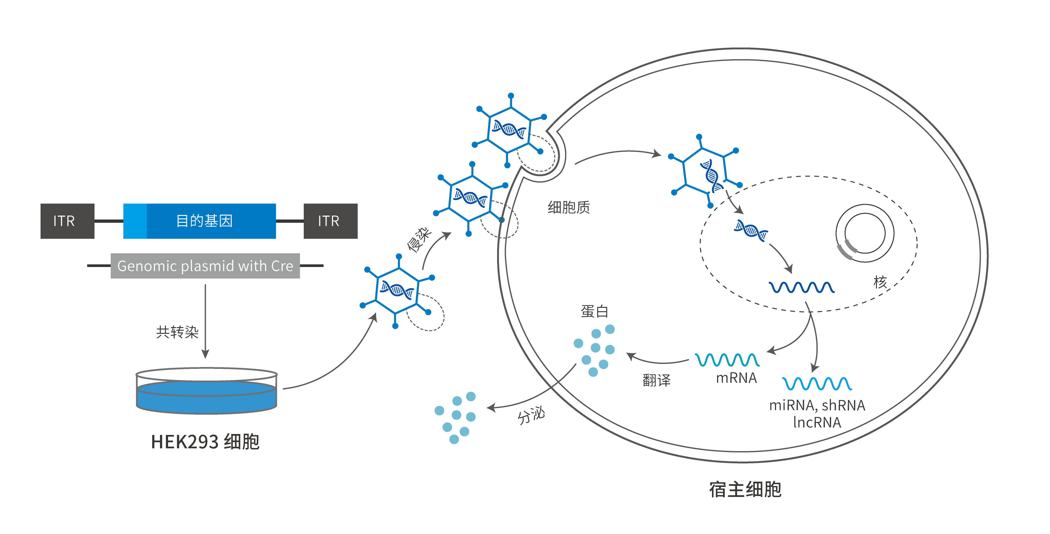
ADV packaging and the infection process
Adenovirus Packaging and Purification
The adenoviral backbone plasmid and the shuttle plasmid are co-transfected into HEK-293 cells. Once cytopathic effects (CPE) become apparent, the viral particles are harvested from both the cell supernatant and the cell lysate, followed by purification via concentration or cesium chloride density-gradient centrifugation.
CPE (Cytopathic Effect): Cellular degeneration resulting from viral infection of cultured cells; this cytopathic effect can be used for quantitative virus assays.
Adenovirus Titer Assay
Assay method: Adenovirus titer determination by enzyme-linked immunosorbent assay; the viral titer is calculated based on the number of positive cells that turn brown after infection.
Experimental principle: In cells positive for adenovirus infection, the capsid proteins are stained brown.
Detection method: Immunochromatographic assay
Experimental principle: Monoclonal antibodies against the capsid protein of adenovirus type 5 are used to bind to cells infected with the adenovirus sample. A HRP‑labeled secondary antibody then binds to the primary antibody, and a chromogenic substrate is added to produce a color change; infected cells turn brown. The viral titer is determined by counting the number of positive cells.
Advantages of Adenoviral Vectors
① Rapid expression: After adenoviral infection, expression can be observed within 1–2 days.
② It has a high vector capacity and high infection efficiency, and is often used to infect cells that are difficult to transduce.
③ Does not integrate into the chromosome and lacks insertional mutagenicity.
④ It exhibits hepatotropism, readily infecting hepatocytes; in vivo applications of adenoviruses are aimed at targeting hepatic infection.
However, due to the high immunogenicity of adenoviruses, they can readily induce inflammation in certain animal models and tissues that are sensitive to exogenous stimuli; therefore, their in vivo application should be approached with caution.
Comparison of the Biological Characteristics of Different Viruses
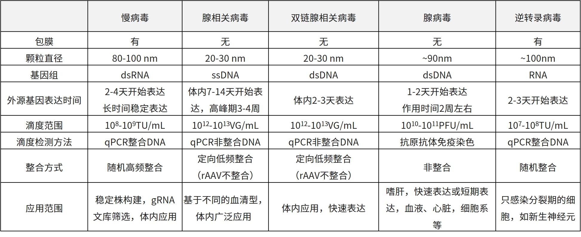
Carrier Selection
HeYuan Bio boasts an extensive portfolio of adenoviral products, suitable for manipulating both coding and non-coding genes, including lncRNAs, miRNAs, and circRNAs.
The following adenoviral vectors are available for your selection:
|
Regulation method |
Carrier Number |
Vector name (vector element order) |
Eukaryotic resistance |
Fluorescence |
Promoter |
Default label |
Carrier capacity |
|
Overexpression |
H225 |
pADV-mCMV-MCS-3xFLAG |
N/A |
N/A |
mCMV |
3FLAG |
>6kb |
|
H201 |
pADV-mCMV-EGFP-3xFLAG |
N/A |
EGFP |
mCMV |
3FLAG |
>5kb |
|
|
H213 |
pADV-mCMV-3xFLAG-EGFP |
N/A |
EGFP |
mCMV |
3FLAG |
>5kb |
|
|
H221 |
pADV-mCMV-3xFLAG-P2A-EGFP |
N/A |
EGFP |
mCMV |
3FLAG |
>5kb |
|
|
H204 |
pADV-mCMV-mCherry-HA |
N/A |
mCherry |
mCMV |
HA |
>5kb |
|
|
H222 |
pADV-mCMV-3xFLAG-T2A-mCherry |
N/A |
mCherry |
mCMV |
3FLAG |
>5kb |
|
|
H12588 |
pADV-EF1-mNeonGreen-CMV-MCS-3xFLAG |
N/A |
mNeonGreen |
CMV |
3FLAG |
>4kb |
|
|
H12589 |
pADV-EF1-mScarlet-CMV-MCS-3xFLAG |
N/A |
mScarlet |
CMV |
3FLAG |
>4kb |
|
|
circRNA overexpression |
H6946 |
pADV-CMV-S-circRNA |
N/A |
N/A |
CMV |
N/A |
>5kb |
|
miRNA overexpression |
H216 |
pADV-U6-miR30(miRNA)-CMV-EGFP |
N/A |
EGFP |
U6 |
N/A |
N/A |
|
Express cre |
K0024 |
pADV-CMV-Cre |
N/A |
N/A |
CMV |
N/A |
N/A |
|
Interference |
DKD001 |
pADV-U6-shRNA-CMV-EGFP |
N/A |
EGFP |
U6 |
N/A |
N/A |
|
DKD004 |
pADV-U6-shRNA-CMV-mCherry |
N/A |
mCherry |
U6 |
N/A |
N/A |
|
|
CRISPR knockout |
H5064 |
pADV-U6-spgRNA v2.0-CMV-3xFLAG-spCas9 |
N/A |
N/A |
U6 |
3FLAG |
N/A |
|
H9350 |
pADV-U6-spgRNA v2.0-CMV-sfGFP-P2A-3xFLAG-spCas9 |
N/A |
sfGFP |
U6 |
3FLAG |
N/A |
|
|
H5066 |
pADV-U6-spgRNA v2.0-CMV-EGFP |
N/A |
EGFP |
U6 |
N/A |
N/A |
|
|
H218 |
pADV-CMV-mCherry-P2A-3xFLAG-spCas9 |
N/A |
mCherry |
CMV |
3FLAG |
N/A |
|
|
H5449 |
pADV-CMV-mCherry-P2A-3xFLAG-eSpCas9_1.1 |
N/A |
mCherry |
CMV |
3FLAG |
N/A |
Application Examples
1. Hepatology. (IF=14.079). Zhou YF, et al. (2018). Cystathionine β-synthase is required for body iron homeostasis. [Adenovirus, Liver]
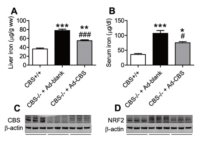
Site of infection: Liver
Vector Name: Ad-CBS
Injection route: tail vein
Injection volume: 1 × 10¹¹ PFU/mL, 10 µL
Testing period: 7 days
2. Diabetes. (IF=8.095). Xiao Y Z, et al. (2015). Activation of ERK1/2 ameliorates liver steatosis in leptin receptor-deficient (db/db) mice via stimulating ATG7-dependent autophagy. [Adenovirus, Liver]
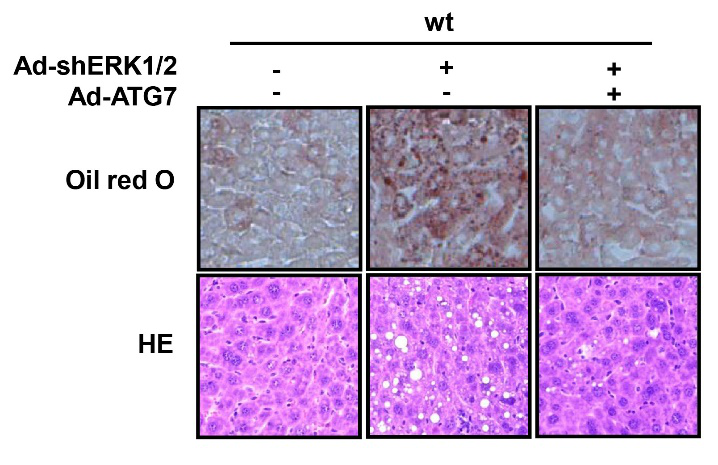
Site of infection: Liver
Vector Name: Ad-ATG7
Injection route: tail vein
Injection dose: 1 × 10^9 PFU per mouse
Testing period: 10 days
3. Molecular Neurobiology. (IF=5.397). Guo J, et al. (2015). Overexpression of Fibulin-5 Attenuates Ischemia/Reperfusion Injury After Middle Cerebral Artery Occlusion in Rats. [Adenovirus, rats]
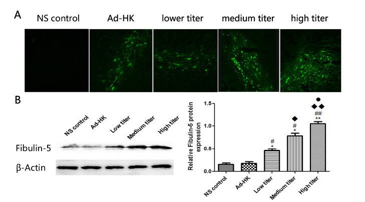
Injection site: rat cerebral cortex
Vector Name: Ad-FBLN
Injection method: stereotactic brain injection
Injection volume: three different doses
Testing period: 7 days
HeYuan Bio employs internationally recognized standard procedures for the production and quality control of adenoviruses, utilizing the AdEasy and Admax adenoviral packaging systems in HEK-293 cells. The resulting viral particles are purified by ultracentrifugation through a density gradient and filtration, with titers determined via capsid protein–based immunoassays. Typically, adenovirus titers range from 10^10 to 10^11 pfu/mL, fully meeting the requirements of diverse experimental applications.
 Telephone consultation


