


 返回
返回
返回

Principle of phalloidin staining (Protocol for using Alexa Fluor 488–labeled phalloidin)
Phalloidin is a bicyclic heptapeptide toxin that selectively binds to filamentous actin (F‑actin) in both animal and plant cells. This unique selectivity makes it an invaluable tool for studying intracellular F‑actin filaments. Under the light microscope, fluorescently labeled phalloidin–dye conjugates can clearly visualize the morphology and distribution of cellular microfilaments, and they are widely employed in biomedical research.
Phalloidin Dyeing principle
Fluorescently labeled phalloidin can selectively bind to F‑actin at the nanogram level and is readily soluble in water, providing a highly convenient labeling method for visualizing and quantifying actin localization in tissue sections, cultured cells, and cell-free systems. Its fluorescent properties enable it to emit light upon excitation at specific wavelengths, thereby rendering the structure and distribution of intracellular actin filaments clearly visible.
Phalloidin Instructions for Use
Taking Alexa Fluor 488–labeled phalloidin (green) as an example.
Step 1: Prepare the 1× Alexa Fluor 488–labeled phalloidin working solution by pipetting 1 μL of the 1000× Alexa Fluor 488–labeled phalloidin stock solution into 1 mL of PBS buffer containing 1% BSA. This will yield a 1× working solution.
Step 2: Cell Staining Procedure
(1) Culture the cells overnight or longer until they reach a confluence of 50–60%.
(2) Aspirate the culture medium, then wash the cells twice with 1× PBS (pH 7.4) pre-warmed to 37°C.
(3) Fix the cells using a PBS solution containing 3–4% formaldehyde, and incubate at room temperature for 10–30 minutes.
(4) At room temperature, wash the fixed cells with PBS 2–3 times, each time for 10 minutes.
(5) (Optional): At room temperature, permeabilize the sample for 3–5 minutes with a 0.1% Triton X-100 solution dissolved in PBS to enhance its permeability.
(6) At room temperature, wash the cells with PBS 2–3 times, 10 minutes each time.
(7) Add 100 μL of Alexa Fluor 488‑labeled phalloidin working solution per well (96-well plate), and incubate at room temperature in the dark for 20–90 minutes; the incubation time may be shorter in summer.
(8) Wash the cells three times with PBS, 5 minutes each time, to remove excess Alexa Fluor 488–labeled phalloidin.
(9) (Optional): Stain the cell nuclei with an adequate volume of ready-to-use DAPI solution, e.g., 100 μL per well (96-well plate), for 3–5 minutes at room temperature. Wash the cells twice with PBS, 5 minutes each time.
(10) Perform fluorescence imaging using a fluorescence microscope or a confocal microscope, selecting either the Alexa Fluor™ 488 excitation/emission filter set and/or the DAPI excitation/emission filter set (Ex/Em = 364/454 nm).
Phalloidin Precautions
✔ Storage conditions: Phallacidin and its fluorescently labeled derivatives should be stored at –20°C or lower, kept dry, and protected from light.
✔ Cell processing: Prior to phalloidin staining, cells should undergo appropriate fixation and permeabilization to ensure optimal staining results.
✔ Fluorescence microscope settings: Adjust the fluorescence microscope settings according to the excitation and emission wavelengths of the fluorescently labeled phalloidin used, to achieve optimal imaging.
✔ Different cell types exhibit varying staining patterns, so the appropriate amount of Alexa Fluor phalloidin should be adjusted accordingly.
✔ Highly toxic; wear gloves when handling.
✔ Avoid fixatives containing methanol, as methanol may disrupt actin during the fixation process.
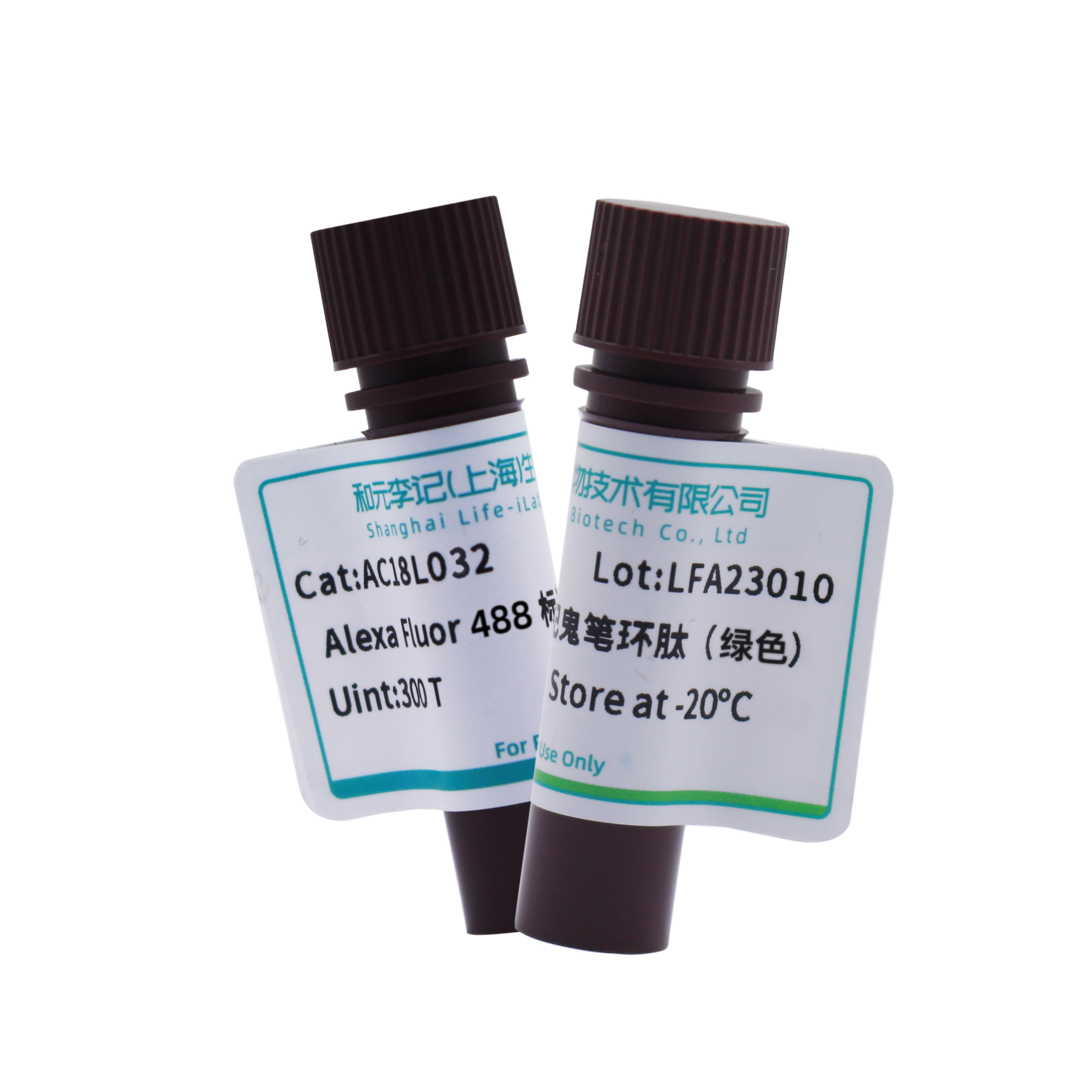
488 green phallotoxin peptide Product Recommendations
[Brand] He Yuan Li Ji
[Product] 488 Phalloidin (green)
[Item Number] AC18L032
[Specification] 300T
Group Website
-
Contact Information
Landline phone:021-50778506
Business Manager:18616108315Business Cooperation/Scholarships:18616108697
Address:4th Floor, Building 27, Lane 908, Ziping Road, International Medical Park, Pudong New Area, Shanghai
Email:ztt0005@obiosh.com

WeChat Official Account
Online Customer Service 1
Online Customer Service 2
 Telephone consultation


