


 返回
返回
返回

A Comprehensive Guide: How to Perform Apoptosis Assays Using the Annexin V–PI Method
The process of apoptosis is often accompanied by distinct morphological changes; for example, in the early stages of apoptosis, phosphatidylserine on the inner leaflet of the plasma membrane translocates to the outer surface. In the mid-to-late phases, as plasma membrane permeability increases, larger molecules such as propidium iodide (PI) can freely diffuse into the cell, resulting in red fluorescence staining of the nucleus.
Principle of the Annexin V–PI Assay
Annexin V is a calcium‑dependent phospholipid‑binding protein that binds to PS with high affinity and specificity. It cannot penetrate intact cell membranes, whereas the membranes of cells undergoing early apoptosis remain intact and are excluded from PI staining. However, in cells at later stages of apoptosis and in dead cells, PI can cross the membrane and stain the nuclei. Therefore, the simultaneous use of Annexin V and PI allows for the discrimination of cells in the early stages of apoptosis from other cell types.
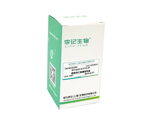
Annexin V–PI Apoptosis Assay Recommendation (Li Ji Bio)
[Name] Flow Cytometry Apoptosis Assay Kit
[Item Number] AC12L033
[Specification] 100T
Annexin V–PI Assay Procedure
▶ Settings for the Control Panel
1. Blank tube: Cells undergoing apoptosis are not stained with the dye and are used to adjust the voltage.
2. Single-stain tubes: Two separate single-stain tubes are required—one containing only PI and the other containing fluorescently labeled Annexin V—used for adjusting compensation.
3. Experimental tube: Cells undergoing apoptosis are treated with all fluorescent dyes used in this experiment.
4. Negative control: Add the dye to a culture of normal cells to confirm their healthy state and rule out false positives.
▶ Suspension cell collection and staining
1. After performing the apoptosis-inducing treatment, centrifuge for 5 minutes, discard the supernatant, collect the cells, gently resuspend them in PBS, and count the cells.
2. Take 1–5 × 10^5 resuspended cells, centrifuge for 5 minutes, discard the supernatant, and add 500 µl of binding buffer to gently resuspend the cells.
3. Add 5 µL of Annexin V-FITC and gently mix; then add 5 µL of propidium iodide and gently mix again.
4. Incubate at room temperature in the dark for 10 minutes. Aluminum foil can be used to block light.
5. Subsequently, flow cytometry was performed, with Annexin V-FITC exhibiting green fluorescence and PI exhibiting red fluorescence.
▶ Adherent cell collection and staining
1. Aspirate the cell culture medium into an appropriate centrifuge tube, wash the adherent cells once with PBS, and then digest the cells with trypsin solution (preferably without EDTA).
2. After the cells have been detached by digestion, follow the same procedure as for suspension cells.
▶ On-site testing
Following staining and incubation, perform flow cytometry or fluorescence microscopy immediately.
Annexin V–PI flow cytometry results (for reference only)
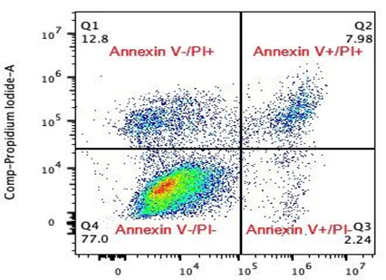
Figure: Apoptosis assay results from a flow cytometer
✔ Upper-left quadrant (Annexin V−/PI+): corresponds to cell debris, mechanically damaged cells, or other dead cells. The greater the number of cells in this quadrant, the lower the reliability of the experimental results; therefore, efforts should be made to minimize the proportion of cells in this quadrant.
✔ Lower-left quadrant (Annexin V-/PI-): represents normal viable cells;
✔ Upper-right quadrant (Annexin V+/PI+): Represents cells in the late stages of apoptosis or necrotic cells; in cells with compromised plasma membranes, DNA can be stained by PI, resulting in red fluorescence.
✔ Lower-right quadrant (Annexin V+/PI-): Represents cells in the early stages of apoptosis; during this phase, PI does not bind to the cells, and no red fluorescence signal is detected.
Precautions for Annexin V–PI Assay
• When working with adherent cells, if it is difficult to precisely control the digestion time, we recommend using Accutase cell dissociation solution, which causes minimal cellular damage and does not alter the distribution of PS on the cell membrane.
• When selecting a fluorescent dye for Annexin V, consider the cell’s intrinsic autofluorescence, particularly in cells that have been cultured for extended periods or treated with drugs. Be especially mindful of the fluorescence emitted by cellular metabolites and the drug itself. In general, the autofluorescence of cellular metabolites can interfere with FITC/PE detection; therefore, Annexin V–APC is often recommended. To assess potential interference from a drug, examine its emission spectrum to determine whether it overlaps with the excitation or emission wavelengths of the dye used in your assay.
• After cell staining is complete, do not fix the samples; perform the assay as soon as possible. The assay tubes may be kept on ice to minimize the impact of external environmental factors on the results.
• In apoptosis assays, it is customary to report the proportion of early-stage apoptosis, as only early apoptosis is biologically meaningful. Of course, some studies also report the total apoptosis rate—defined as the sum of early and late-stage apoptosis—though reporting the proportion of late-stage apoptosis alone is of limited interpretive value.
Group Website
-
Contact Information
Landline phone:021-50778506
Business Manager:18616108315Business Cooperation/Scholarships:18616108697
Address:4th Floor, Building 27, Lane 908, Ziping Road, International Medical Park, Pudong New Area, Shanghai
Email:ztt0005@obiosh.com

WeChat Official Account
Online Customer Service 1
Online Customer Service 2
 Telephone consultation


