


 返回
返回
返回

Principles and Procedures for Detecting Reactive Oxygen Species (ROS)
Reactive oxygen species (ROS) are oxidatively active chemical compounds generated within cells, including hydrogen peroxide and the superoxide anion. ROS are produced under normal physiological conditions; however, when their production exceeds the cell’s capacity to detoxify them, oxidative stress ensues, leading to cellular damage and disease.
Detection principle
The most widely used method for detecting intracellular ROS is the DCFH-DA fluorescence probe assay. DCFH‑DA (2′,7′‑dichlorofluorescein diacetate) is a fluorescent probe that is nonfluorescent in its native form but can freely cross the cell membrane. Once inside the cell, it is hydrolyzed by intracellular esterases into DCFH. Because DCFH cannot pass through the cell membrane, the fluorescent probe accumulates within the cell. Intracellular ROS can oxidize the nonfluorescent DCFH, generating fluorescent DCF; the fluorescence intensity is directly proportional to the ROS level. Fluorescence is detected using instruments such as a fluorescence microscope, flow cytometer, or laser scanning confocal microscope, with excitation at 480 nm and emission at 525 nm.
Testing steps
I. Loading Probe
Cells subjected to short‑duration stimulation (typically ≤2 h): first load the probe, then stimulate the cells with an ROS‑positive control or with a compound of interest.
Cells subjected to prolonged stimulation (typically for 6 hours or longer): first stimulate the cells with an ROS-positive control or with a compound of interest, then load the probe.
1. In situ probe loading (applicable only to adherent cells)
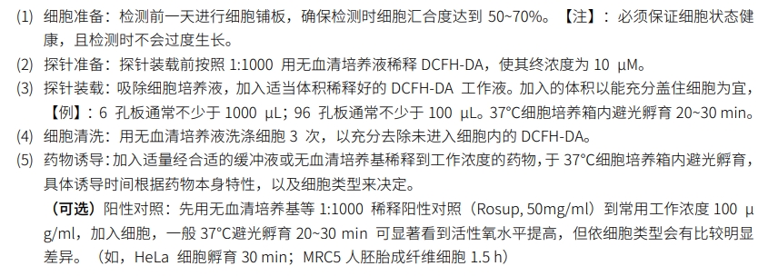
2. Load the probe after cell collection (suitable for both adherent and suspension cells).
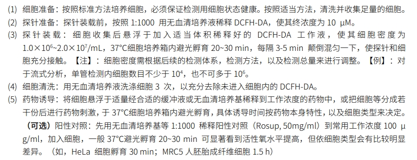
II. Testing
In situ probe‑loading method: direct observation by laser confocal microscopy, or, after cell collection, detection using a fluorescence spectrophotometer, a fluorescence microplate reader, or a flow cytometer.
After cell collection, load the probes: detection can be performed using a fluorescence spectrophotometer, a fluorescence microplate reader, or a flow cytometer; alternatively, direct observation is possible with a laser confocal microscope.
Precautions
① After probe loading, be sure to thoroughly wash away any residual probes that have not entered the cells; otherwise, background fluorescence will be elevated.
② After the probe has been loaded and any residual probes have been washed away, you can perform scans of the excitation wavelength and the emission wavelength to verify whether the probe loading is satisfactory.
Good.
③ Minimize the time between probe loading and measurement (excluding the stimulation period) to reduce potential sources of error.
④ Based on the overall fluorescence intensity observed in the results, the dilution factor of the DCFH‑DA probe and the incubation time can be adjusted accordingly.
Group Website
-
Contact Information
Landline phone:021-50778506
Business Manager:18616108315Business Cooperation/Scholarships:18616108697
Address:4th Floor, Building 27, Lane 908, Ziping Road, International Medical Park, Pudong New Area, Shanghai
Email:ztt0005@obiosh.com

WeChat Official Account
Online Customer Service 1
Online Customer Service 2
 Telephone consultation


