


 返回
返回
返回


Reactive Oxygen Species (ROS) Assay Kit

One click copying of product information
-
AC12L925100T480
- Product Description
- Product FAQ
- COA
- Product Literature
- MSDS
- Operation video
-
Product Description
Reactive oxygen species (ROS), including the superoxide anion, hydrogen peroxide, and their downstream products such as peroxides and hydroxyl radicals, are involved in cellular growth and proliferation, development and differentiation, aging and apoptosis, as well as numerous physiological and pathological processes. Meilun’s Reactive Oxygen Species Assay Kit is the most widely used method for quantitatively measuring intracellular ROS levels, based on changes in fluorescence intensity of the fluorescent dye DCFH-DA (7-Dichlorodihydrofluorescein diacetate).
DCFH‑DA itself is nonfluorescent and can freely cross the cell membrane. Once inside the cell, it is hydrolyzed by intracellular esterases to yield DCFH. However, DCFH cannot permeate the cell membrane, allowing the probe to be readily retained within the cell. In the presence of reactive oxygen species (ROS), DCFH is oxidized to produce the fluorescent compound DCF; the intensity of the green fluorescence is directly proportional to the intracellular ROS level. By measuring the fluorescence of DCF, one can determine the cellular ROS levels. Using a fluorescence microscope, laser scanning confocal microscope, fluorometer, fluorescence microplate reader, or flow cytometer, DCF fluorescence can be detected at an excitation wavelength of 502 nm and an emission wavelength around 530 nm, thereby quantifying intracellular ROS levels.
This kit features low background, high sensitivity, a wide linear range, and ease of use, making it suitable for detecting reactive oxygen species in various eukaryotic cultured cells. It also includes Rosup, a positive control reagent for ROS, to facilitate ROS detection. Taking a 6-well plate as an example, with 1 mL per well, the kit can perform approximately 100 assays; on a 96-well plate, it supports about 1,000 assays (100 μL per well).
Order Information
Product Name
Item number
Specifications
Reactive Oxygen Species (ROS) Assay Kit
AC12L925
100T
Product ingredients
Component
Specifications
A. DCFH-DA (10mM)
0.1 mL
B. Reactive oxygen species positive control (Rosup, 50 mg/mL)
1 mL
Transportation and Storage
Blue ice transport. Store at -20°C away from light; shelf life: 12 months.
Instructions for Use
I. Loading Probe
Cells subjected to short‑duration stimulation (typically ≤2 h): first load the probe, then stimulate the cells with an ROS‑positive control or with a compound of interest.
Cells subjected to prolonged stimulation (typically for 6 hours or longer): first stimulate the cells with an ROS-positive control or with a compound of interest, then load the probe.
1. In situ probe loading (applicable only to adherent cells)
(1) Cell preparation: Plate the cells the day before the assay to ensure that, at the time of testing, cell confluence is 50–70%. [Note]: The cells must be in good condition and should not exhibit excessive growth during the assay.
(2) Probe preparation: Prior to probe loading, dilute DCFH-DA in serum-free culture medium at a 1:1000 ratio to achieve a final concentration of 10 μM.
(3) Probe loading: Aspirate the cell culture medium and add an appropriate volume of freshly diluted DCFH‑DA working solution. The volume should be sufficient to fully cover the cells; for example, in a 6-well plate, use at least 1,000 μL, and in a 96-well plate, use at least 100 μL. Incubate in a 37°C cell culture incubator, protected from light, for 20–30 minutes.
(4) Cell washing: Wash the cells three times with serum-free culture medium to thoroughly remove any DCFH-DA that has not entered the cells.
(5) Drug induction: Add an appropriate amount of the drug, diluted to the working concentration in a suitable buffer or serum-free medium, and incubate in the 37°C cell culture incubator away from light. The specific induction time should be determined based on the drug’s intrinsic properties and the cell type.
(5) (Optional) Positive control: First dilute the positive control (Rosup, 50 mg/mL) 1:1000 in serum-free medium to obtain a working concentration of 100 μg/mL. Add the cells and incubate at 37°C in the dark for 20–30 minutes; this will typically result in a noticeable increase in reactive oxygen species levels, though the extent of this effect may vary considerably depending on the cell type. For example, HeLa cells should be incubated for 30 minutes, while human embryonic fibroblasts (MRC5) require 1.5 hours.
2. Load the probe after cell collection (suitable for both adherent and suspension cells).
(1) Cell preparation: Cells should be cultured according to standard protocols, ensuring that the cells used for assay are in good condition. Wash and harvest an adequate number of cells using an appropriate method.
(2) Probe preparation: Before loading the probe, dilute DCFH-DA in serum-free culture medium at a 1:1000 ratio to achieve a final concentration of 10 μM.
(3) Probe loading: After cell collection, resuspend the cells in an appropriately diluted DCFH‑DA working solution to achieve a cell density of 1.0 × 10^6 to 2.0 × 10^7 cells/mL. Incubate in a 37°C incubator, protected from light, for 20–30 minutes, gently inverting and mixing every 3–5 minutes to ensure thorough contact between the probe and the cells. [Note]: The cell density should be adjusted according to the downstream assay system, the detection method, and the total amount of sample to be analyzed. [Example]: For flow cytometry, the number of cells per well should be no less than 10^4 and no more than 10^6.
(4) Cell washing: Wash the cells three times with serum-free culture medium to thoroughly remove any DCFH-DA that has not entered the cells.
(5) Drug-induced differentiation: Suspend the cells in an appropriate volume of drug solution diluted to the working concentration using a suitable buffer or serum-free medium, or divide the cells into several aliquots and then expose them to the drug. Incubate the cells in a 37°C cell culture incubator in the dark; the specific induction duration should be determined based on the intrinsic properties of the drug and the cell type.
(5) (Optional) Positive control: First dilute the positive control (Rosup, 50 mg/mL) 1:1000 in serum-free medium to obtain a working concentration of 100 μg/mL. Add the cells and incubate at 37°C in the dark for 20–30 minutes; this will typically result in a noticeable increase in reactive oxygen species levels, though the extent of this effect may vary considerably depending on the cell type. For example, HeLa cells should be incubated for 30 minutes, while human embryonic fibroblasts (MRC5) require 1.5 hours.
II. Testing
In situ probe‑loading method: direct observation by laser confocal microscopy, or, after cell collection, detection using a fluorescence spectrophotometer, a fluorescence microplate reader, or a flow cytometer.
After cell collection, load the probes: detection can be performed using a fluorescence spectrophotometer, a fluorescence microplate reader, or a flow cytometer; alternatively, direct observation is possible with a laser confocal microscope.
III. Parameter Settings
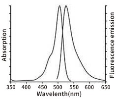
Using an excitation wavelength of 488 nm and an emission wavelength of 525 nm, the fluorescence intensity is monitored in real time or at discrete time points before and after stimulation. The fluorescence spectrum of DCF is very similar to that of FITC, so DCF can be detected using the same parameter settings as FITC. The excitation and emission spectra of DCF are shown in the figure below.
IV. Explanations of Other Matters
1. The positive control Rosup is typically used at a 1:1000 dilution. A significant increase in reactive oxygen species levels can usually be observed 20–30 minutes after stimulation. The effectiveness of the ROS-positive control may vary considerably among different cell types. If no rise in ROS is detected within 30 minutes post‑stimulation, consider extending the incubation period or slightly increasing the concentration of the ROS-positive control. Conversely, if ROS levels rise too rapidly, shorten the incubation time or reduce the concentration of the ROS-positive control accordingly.
2. For certain cell types, if the negative control cells exhibit relatively strong fluorescence even in the absence of stimulation, DCFH‑DA can be diluted at a ratio of 1:2000 to 1:5000, resulting in a final probe concentration of 2–5 μM during loading. The incubation time for probe loading may also be adjusted as needed, ranging from 15 to 60 minutes.
3. The reactive oxygen species positive control (Rosup) is intended solely as a positive control sample and does not need to be added to every sample.
Precautions
1. This product is intended solely for scientific research and must not be used for clinical diagnosis, treatment, or any other such purposes.
2. After probe loading, be sure to thoroughly wash away any residual probes that have not entered the cells; otherwise, background fluorescence will be elevated.
3. After the probe has been loaded and any residual probe has been washed away, you can perform scans of the excitation and emission wavelengths to verify that the probe has been properly loaded. For the excitation and emission spectra of DCF, please refer to the figures on the previous page.
4. Minimize the time between probe loading and assay measurement (excluding the stimulation period) to reduce potential sources of error.
This product is intended solely for scientific research and must not be used for clinical diagnosis, treatment, or any other purposes.
-
Fill in the product batch number for inquiry
-
Fill in the product batch number for inquiry
Best Sellers

Popular Articles
Group Website
-
Contact Information
Landline phone:021-50778506
Business Manager:18616108315Business Cooperation/Scholarships:18616108697
Address:4th Floor, Building 27, Lane 908, Ziping Road, International Medical Park, Pudong New Area, Shanghai
Email:ztt0005@obiosh.com

WeChat Official Account
Online Customer Service 1
Online Customer Service 2
 Telephone consultation


